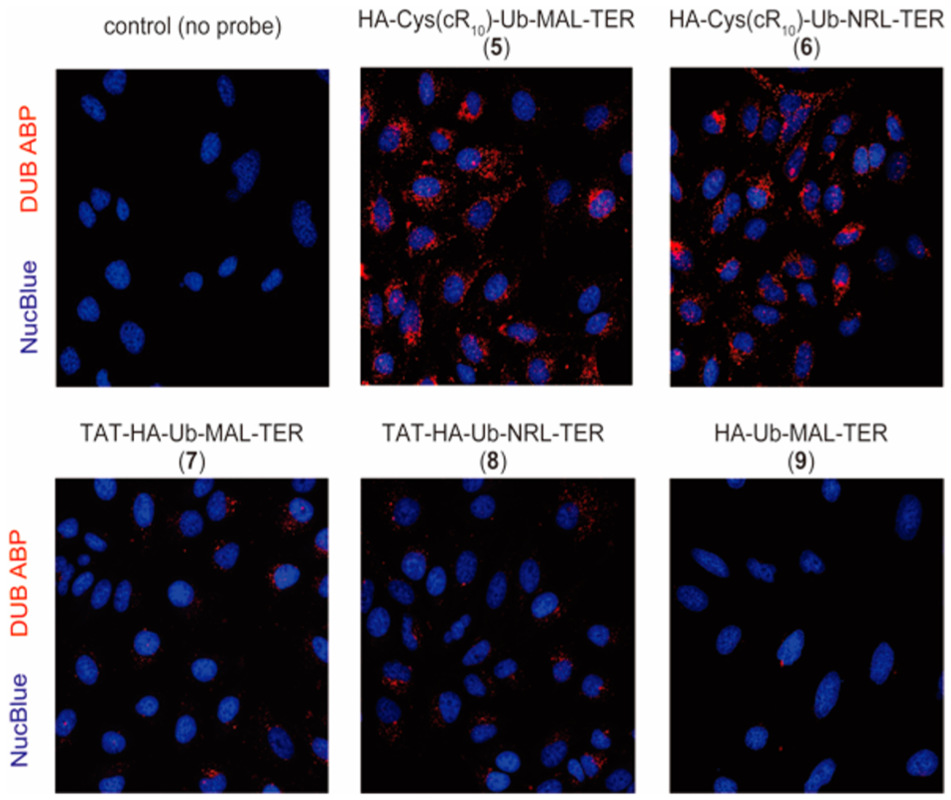
Figure 4.

Live-cell fluorescence imaging of HeLa cells treated with the cell-permeable fluorescent Ub-based DUB ABPs. Comparison of HeLa cells treated with indicated probes 5, 6, 7, 8, and 9 (15 μM, 4 h). DUB ABPs are visualized using TER fluorescence (red channel), and NucBlue Live Readyprobes Reagent was utilized for nuclear staining (blue channel).
