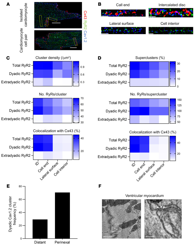
Figure 4. Cx43 colocalizes with large dyadic RyR2 superclusters and forms microdomains at the perinexus.
(A) 2D SMLM images of a murine cardiomyocyte (top) and cardiomyocyte cell pair (bottom), triple stained for Cx43 (red), RyR2 (green), and Cav1.2 (blue). Scale bar: 10 μm. (B) Straightened region of interest (from yellow boxes in A) of Cx43, RyR2, and Cav1.2 at different subcellular domains. Scale bar: 2 μm. (C) Heatmap of RyR2 cluster density, number of molecules, and colocalization with Cx43 at different subcellular domains (n = 5, n = 42 single cardiomyocytes, 16 cardiomyocyte cell pairs). RyR2 clusters were classified as dyadic or extradyadic based on the proximity of Cav1.2 clusters, RyR2 clusters occurring less than 250 nm from a Cav1.2 cluster were categorized as dyadic. (D) Heatmap of RyR2 supercluster abundance, size, and colocalization with Cx43 at different subcellular domains (n = 5, n = 42 single cardiomyocytes, 16 cardiomyocyte cell pairs). (E) Relative localization overview in left ventricular mouse cardiomyocyte cell pairs. Dyadic Cav1.2 clusters were categorized as perinexal or distant based on edge distance 200 nm or less or greater than 200 nm from edge of Cx43 cluster, respectively (n = 5, n = 16 cardiomyocyte cell pairs). (F) EM images of an SR cistern forming a dyadic cleft at the perinexus in mouse ventricular myocardium. Left image shows an EM overview of a murine ventricular intercalated disc. Scale bar: 500 nm. White box is enlarged on the right. Arrows indicate electron dense particles, likely ryanodine receptors. Scale bars: 100 nm. Pn, perinexus.

