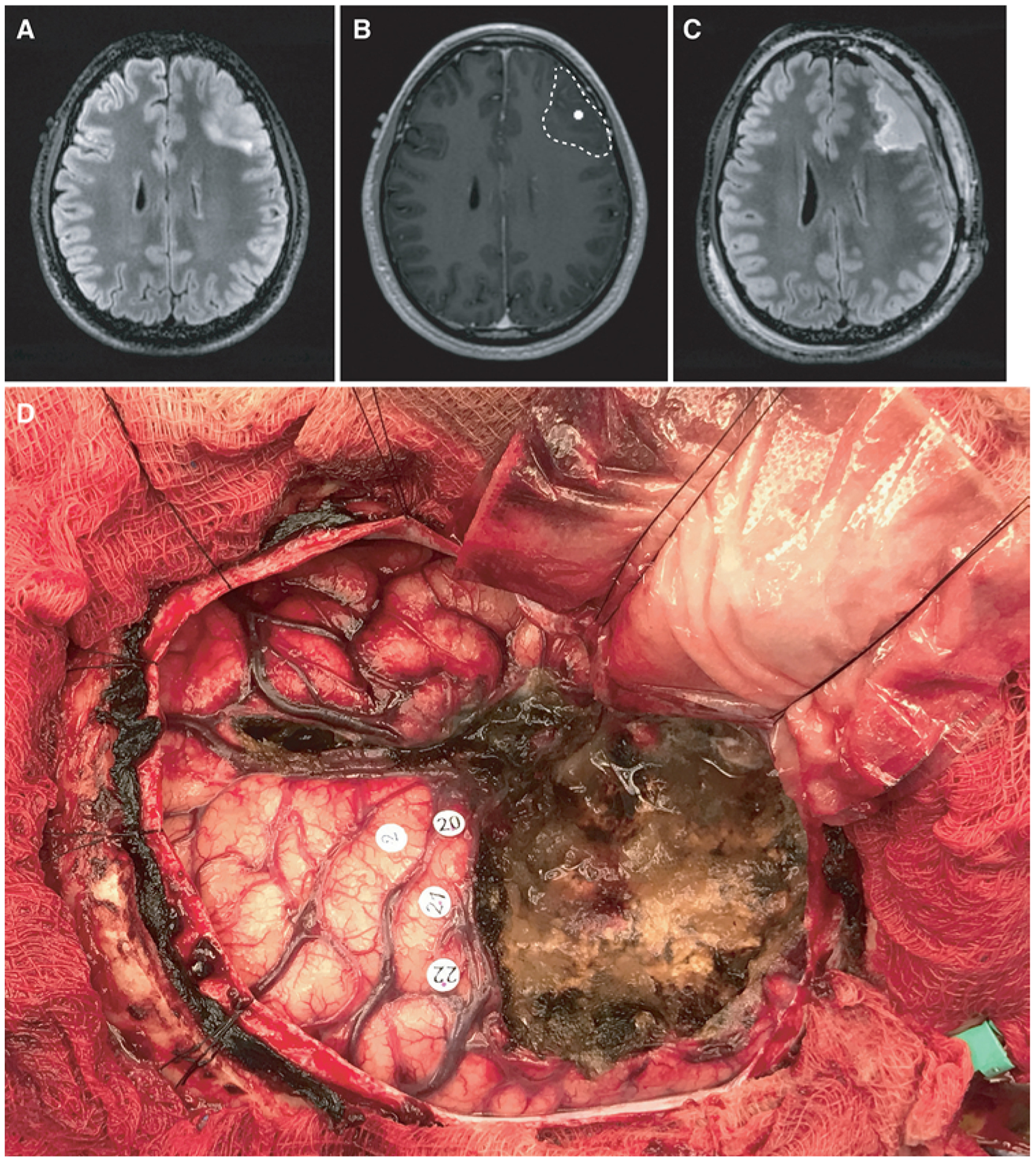
FIG. 5.

An illustrative case. A 41-year-old right-handed man with a left frontal-temporal anaplastic oligodendroglioma status after he underwent a debulking procedure at an outside hospital. The patient had no preoperative language deficits. A: Preoperative T2-weighted FLAIR MR image revealing an infiltrative T2 hyperintensity within the left anterior frontal lobe. B: Preoperative MEG image of an HFC hub superimposed on the preoperative T1-weighted post-Gd MR image. The tumor margin is demarcated by the hashed line. C: Postoperative T2-weighted FLAIR image showing a 95% extent of resection. D: Intraoperative photograph taken after tumor resection. Number 2 represents the face motor cortex. Numbers 20, 21, and 22 mark anomia sites identified by IES.
