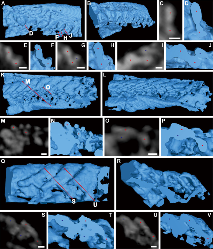
Fig. 2. Dumbbell-shaped filaments of T. eatoni.
(A to J) Computed tomography (CT) reconstruction of partial limb, USNM 65527. (A) Dorsal view. (B) Posterolateral view. (C to J) Cross sections of filaments in (A) showing dumbbell-shaped outline with inflated marginal bulbs connected by narrow central region. (K to V) CT reconstruction of partial limb, USNM 65523. (K) Dorsal view. (L) Posterolateral view. (M to P) Cross sections of filaments in (K) showing separately preserved marginal bulbs. (Q) Dorsal view. (R) Posterior view. (S to V) Cross sections of filaments in (Q) show inflated marginal bulbs connected by narrow central region. Asterisks locate the top and bottom inflated marginal bulbs of dumbbell-shaped filaments, which are connected by a narrow central region. CT reconstructions are shown as blue color. CT slices are displayed as gray color. The same color of asterisks represents the same filament. Red lines represent the position of cross sections. Scale bars, 30 μm.

