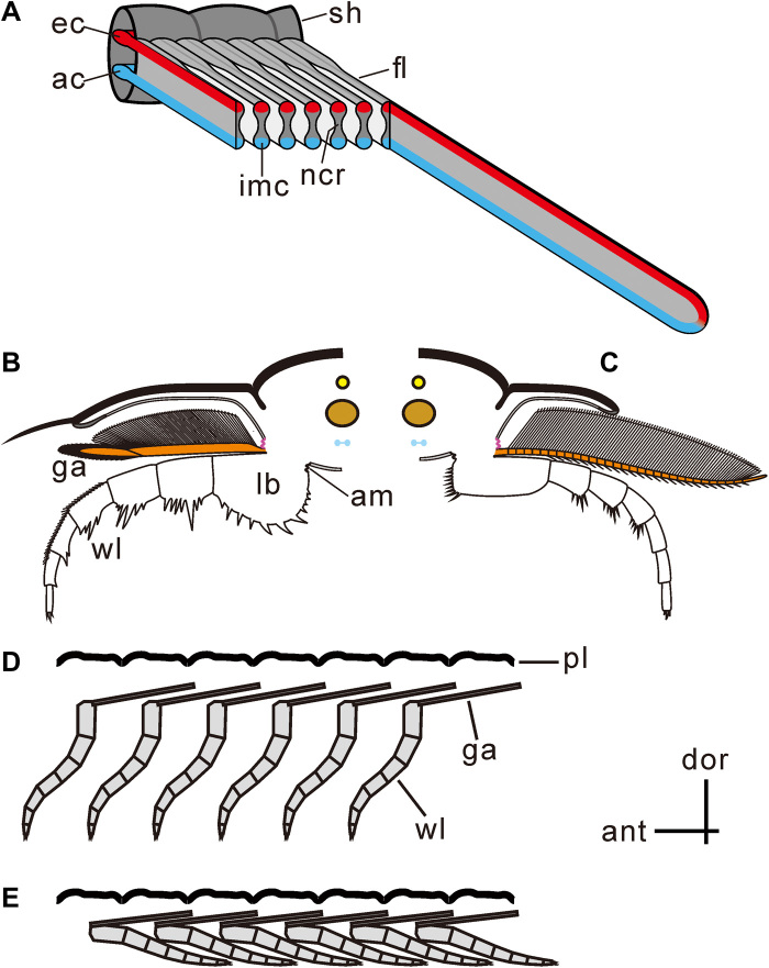
Fig. 4. Reconstructions of trilobite limbs.
(A) Filaments showing dumbbell-shaped cross section with inflated marginal bulbs and narrow central region, of which the inflated marginal bulbs provide afferent and efferent channels and the narrow central region functioning for respiratory exchange. (B and C) Cross section showing the upper branch connecting dorsally with the extended arthrodial membrane (purple) and ventrally with the proximal limb base. The upper limb extended posterodorsally, as shown in Fig. 6. (B) Proposed articulation of O. serratus [modified from the work of Ramsköld and Edgecombe (41)]; anterior view of right limb. (C) Proposed articulation of T. eatoni [modified from the work of Whittington and Almond (57)]; anterior view of left limb. (D and E) Imbrication style of two branches limited the movement of the upper branch. (D) Upper branch showing anterior imbrication style, resulting from limited space for limb swinging. (E) Upper branches preserved in anterior imbrication style with lower branches preserved in posterior imbrication style. am, extended arthrodial membrane; ant, anterior; ac, afferent channel; dor, dorsal; ec, efferent channel; fl, filament; ga, gill appendage; imc, inflated marginal channel; lb., limb base; pl, pleura; ncr, narrow central region; sh, shaft; wl, walking leg.

