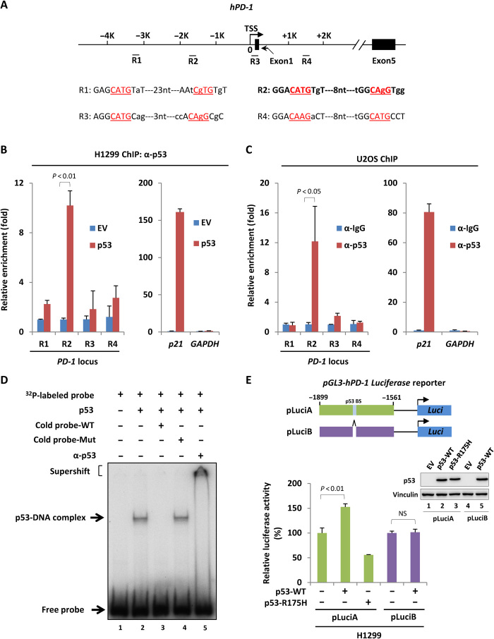
Fig. 2. Identification of PD-1 as a p53 direct target gene.
(A) Schematic diagram of human PD-1 gene locus with four potential p53-binding regions. TSS, transcription start site. (B) ChIP-qPCR analysis of p53 occupancy on PD-1 promoter or gene body region in H1299 cells transfected with empty vector or p53-expressing construct for 24 hours. (C) ChIP-qPCR analysis of p53 occupancy on PD-1 promoter or gene body region in U2OS cells. p53-binding on p21 and GAPDH promoter in (B) and (C) was measured as positive and negative control, respectively. (D) EMSA of p53 binding with PD-1 promoter in vitro. Purified p53 was incubated with a 32P-labeled probe containing p53-binding element of PD-1 promoter. α-p53 antibody was used for supershift assay. (E) Luciferase assay of p53-driven transcription of PD-1 promoter–containing reporter expressing cassette in H1299 cells. The expression of p53 was detected by Western blot. BS, binding site. Data were shown as means ± SD, n = 3.

