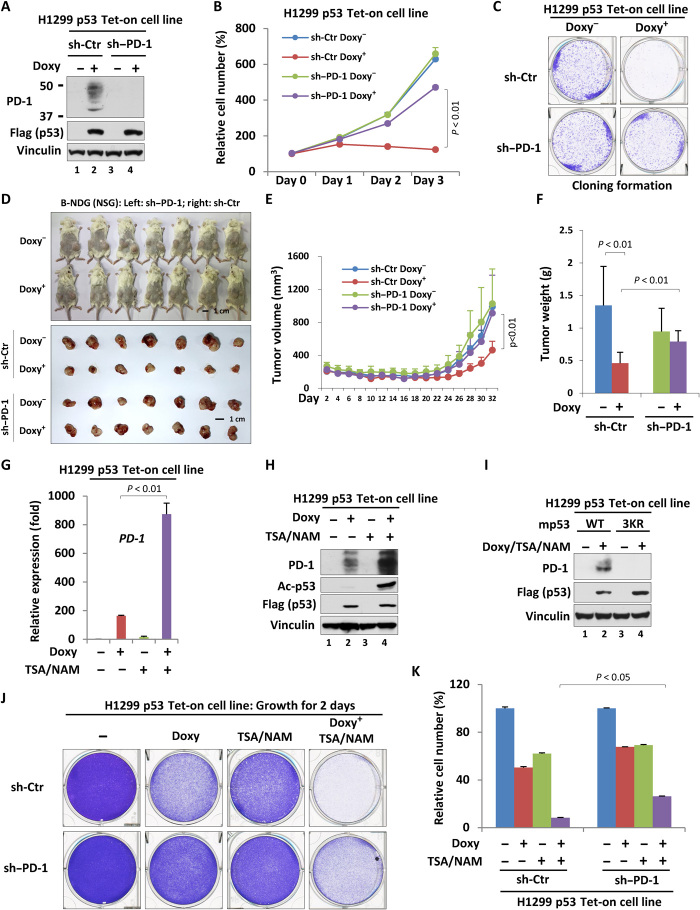
Fig. 6. Activation of cancer cell–intrinsic PD-1 participates in p53-dependent tumor suppression.
(A) Western blot analysis of PD-1 knockdown efficiency in the H1299 p53 Tet-on cell line. (B) Cell proliferation assay for three consecutive days of H1299 p53 Tet-on control or PD-1–depleted cells treated with or without doxycycline. (C) Cloning formation of H1299 p53 Tet-on control or PD-1 depleted cells treated with or without doxycycline. (D) Xenograft tumor growth analysis of PD-1 effects on p53-mediated tumor suppression in B-NDG (NSG) mice. (E) The volume analysis of xenograft tumors based on (D). (F) The final weight of xenograft tumors based on (D). (G and H) RT-qPCR (G) or Western blot (H) analysis of the mRNA or protein level of PD-1 in H1299 p53 Tet-on cells treated with doxycycline (for 24 hours) and/or TSA/NAM (for the last 6 hours). (I) Western blot analysis of PD-1 in H1299 p53 Tet-on (wild-type versus 3KR) cells treated with or without doxycycline (for 24 hours) and TSA/NAM (for the last 6 hours). (J) Cell proliferation analysis (48 hours) of H1299 p53 Tet-on control or PD-1–depleted cells treated with or without doxycycline. (K) Quantitation of cell proliferation based on (J). Data were shown as means ± SD, n = 3 or 7. Photo credit: Zhijie Cao, Chinese Academy of Medical Sciences and Peking Union Medical College.

