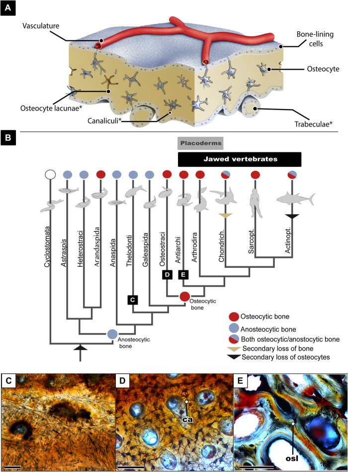
Fig. 1. Overview of the evolution of osteocytes.
(A) Schematic drawing showing bone microanatomy; *denotes the bone anatomy that is readily preserved in fossil material. (B) Vertebrate phylogeny showing the evolution of osteocytic versus anosteocytic bone modified from (38). (C) Image of histological section of fossilized anosteocytic bone of a heterostracan (MB.f.TS.2302) showing tubules but no lacunae present. (D) Histological section of fossilized osteocytic bone of an the osteostracan Tremataspis mammillata (MB.f.TS.463) showing dense osteocyte lacunae and canaliculi. (E) Histological section of fossilized placoderm bone showing both osteocyte lacunae and osteonal remodeling. osl, osteocyte lacunae; ca, canaliculi. All scale bars are 100 μm.

