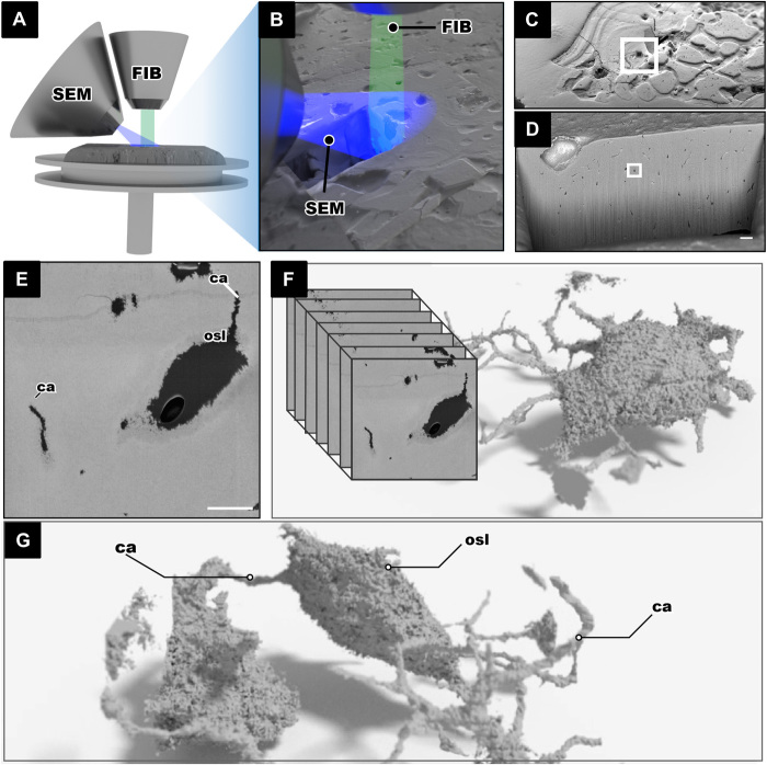
Fig. 3. FIB-SEM tomography imaging and processing of the fossil jawless vertebrate T. mammillata (MB.f.9025).
(A and B) FIB-SEM setup showing the FIB in relation to the SEM both aimed at the region of interest. (C) Bone surface with an excavate area made by the FIB. (D) Internal wall of the excavated area lined with small black dots that are the fossil osteocyte lacunae. (E) Single osteocyte lacuna from the surface that is scanned; the single SEM image shows the lacunae and canaliculi in black and the mineralized bone in gray; scale bar, 5 μm. (F and G) An image stack is obtained, and 3D made of fossil LCN can be made.

