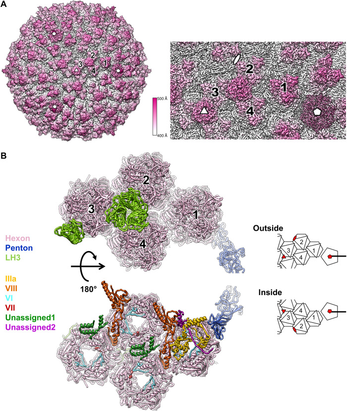
Fig. 1. LAdV-2 cryo-EM map and molecular model.
(A) Right: Surface rendering of the LAdV-2 three-dimensional (3D) map colored by radius from white to pink, as indicated by the color scale. A zoom-in on the area corresponding to the icosahedral asymmetric unit (AU) is shown at the right. The four hexon trimers in an AU are numbered 1 to 4. White symbols indicate the icosahedral fivefold (pentagon), threefold (triangle), and twofold (oval) symmetry axes. (B) Ribbon representation of the proteins traced in the AU, colored as indicated by the legend at the left. Depth cueing (fading to white) is used to give an impression of distance to the viewer. Two views are provided, as seen from outside (top) or inside (bottom) the capsid, with a cartoon at the right-hand side for guidance. In the cartoon, icosahedral symmetry axes are indicated by red symbols.

