Figure 6. Syt-7 stimulates PMA-induced potentiation of release at low prestimulation [Ca2+].
(A) Calcium uncaging experiment from low prestimulation [Ca2+] in Syt-7 WT cells (persian green) and in Syt-7 WT cells perfused with 100 nM phorbol 12-myristate 13-acetate (PMA) (WT + PMA) (gray). Panels are arranged as in Figure 1A. PMA treatment strongly augmented the primed pool size in WT cells. (B) Sizes of the RRP and SRP. (C) Time constants, τ, of fusion for fast (i.e. RRP) and slow (i.e. SRP) secretion. (D) Sustained rates of secretion. (E) Calcium uncaging experiment from low prestimulation [Ca2+] in Syt-7 KO cells (vermilion) and in Syt-7 KO cells perfused with 100 nM PMA (KO + PMA) (gray). PMA-induced potentiation of release was much weaker in Syt-7 KO cells. (F) Size of the RRP and SRP. (G) Time constants, τ, of fusion for fast (i.e. RRP) and slow (i.e. SRP) secretion. (H) Sustained rate of secretion. Data information: Data with error bars (A–H) are presented as mean ± SEM; in (A, E), the traces are the average of all cells. Statistics: *: p<0.05; **p<0.01; ***p<0.001; ****p<0.0001. Analysis was performed with Mann-Whitney test. Number of cells: WT: N = 23 cells; WT + PMA: N = 18 cells; Syt-7 KO: N = 22 cells; Syt-7 KO + PMA = 24 cells.

Figure 6—figure supplement 1. Integrated amperometry (mean ± SEM) of WT, WT cells treated with 100 nM phorbol 12-myristate 13-acetate (PMA) (WT + PMA), Syt-7 KO and Syt-7 KO with 100 nM PMA (Syt-7 KO + PMA) stimulated from low prestimulation [Ca2+].

Figure 6—figure supplement 2. Application of phorbol esters to Syt-7 WT and KO at higher prestimulation [Ca2+].

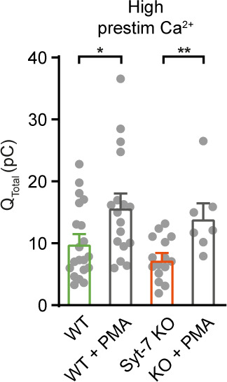
Figure 6—figure supplement 3. Integrated amperometry (mean ± SEM) of WT, WT cells perfused with 100 nM phorbol 12-myristate 13-acetate (PMA) (WT + PMA), Syt-7 KO and Syt-7 KO treated with 100 nM PMA (syt-7 KO + PMA) stimulated from high prestimulation [Ca2+].