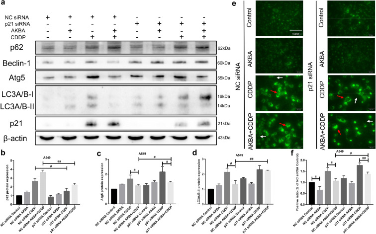
Fig. 11.
AKBA enhanced the sensitivity of CDDP (AKBA 10 μg/ml, CDDP 2 μg/ml) through autophagy suppression via p21-dependent signaling pathway in A549 cells. a p62, Atg5, Beclin-1, LC3A/B, and p21 protein expressions were measured by western blotting assay with or without knockdown of p21 in A549 cells treated with AKBA, CDDP alone, or in combination. b, c, d Histogram showing the level of p62, Atg5, and LC3A/B-II proteins and relative statistical analysis. e Representative images of the formation of autolysosome measured by immunofluorescence in A549. White arrow indicates negative autophagosome cell and the red indicates positive autophagsome cell. Scale bar = 50 μM. f Quantification of the positive phagosomes in A549. Data were represented as the mean ± SD of 3 independent experiments

