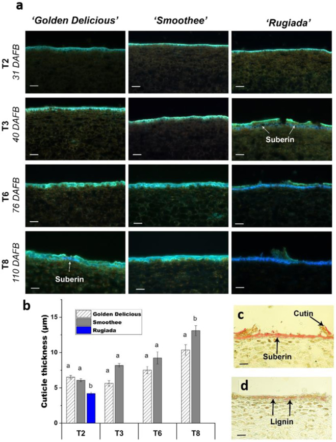
Fig. 2. Light microscopy analysis of the epidermal cell layer of the three apple genotypes.
a Cross-sections of the epidermal layer of ‘Golden Delicious’ and its russet and non-russet clones, ‘Rugiada’ and ‘Smoothee’, showing autofluorescent structures. Flavonoids are mainly responsible for the fluorescence of the cuticle (green). The presence of suberin and/or lignin (blue emission) appears from T3 in ‘Rugiada’ and T8 in ‘Golden Delicious’. Excitation 355 nm with emission at 400–800 nm. Scale bar = 50 µm. b Cuticle thickness measured using the lipid stain Sudan IV. The cuticle of the russet clone ‘Rugiada’, although intact at T2, shows a significantly reduced thickness as compared to ‘Golden Delicious’ and ‘Smoothee’. Significance was calculated according to a one-way ANOVA of p < 0.05. The cuticle of ‘Rugiada’ could not be measured after T2. c Light microscopy of the epidermal layer of T6 stage fruit from ‘Rugiada’ using the lipid stain Sudan IV shows the presence of suberin in the periderm and the dramatic reduction in cuticle deposition (with only patches of cutin remaining). d Phloroglucinol staining of the T8 stage fruit from ‘Rugiada’ showing lignified cell-wall tissue in the periderm (pink and red)

