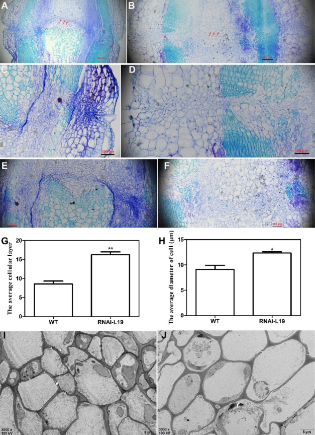
Fig. 3. Anatomic analysis of the fruit pedicel abscission zone of the SlBL4 RNAi plant.
A–F Cross-sections of the fruit pedicel abscission zone at the 25 dpa stage as revealed by toluidine blue staining. A, C, E WT; B, D, and F SlBL4 RNAi-L19. G, H The epidermal cell layers per cell in the fruit pedicel abscission zone of 25 dpa fruit pedicels in WT and SlBL4 RNAi-L19 plates; H The epidermal diameter per cell in the fruit pedicel abscission zone of 25 dpa fruit pedicels in WT and SlBL4 RNAi-L19 plants. I, J Transmission electron micrographs of the fruit pedicels of SlBL4 RNAi-L19 and WT tomato at 25 dpa; I WT; J SlBL4 RNAi-L19; scale bars: 5 μm; dpa day post anthesis, WT wild type

