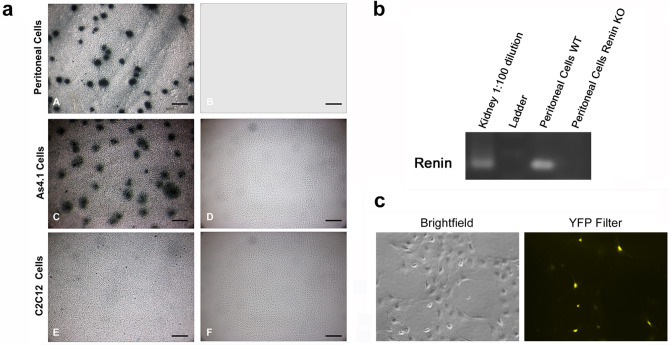
Figure 2.
Peritoneal cells express renin. (a) A dot membrane immunoassay shows that peritoneal cells derived from the renin lineage appear as dark blue dots, indicating that these cells actively manufacture renin (A). A similar pattern is obtained with As4.1 cells, a mouse tumoral cell line that manufactures renin constitutively (C). By the contrary, no spots were detected in C2C12 cells, skeletal muscle cells that do not normally synthesize renin (E). Further, no spots are detected in any of the cells when the membrane immunoassay is performed in the absence of the primary renin antibody (B, D, F) (scale bars A-F 200 µm). (b) Semi-quantitative RT-PCR was performed on wildtype peritoneal cells and peritoneal cells from a renin KO animal. Kidney RNA was used as a positive control. (c) Peritoneal cells from Ren1c-YFP reporter mice, where YFP marks active renin expression were grown in culture. YFP was demonstrated by immunofluorescence.

