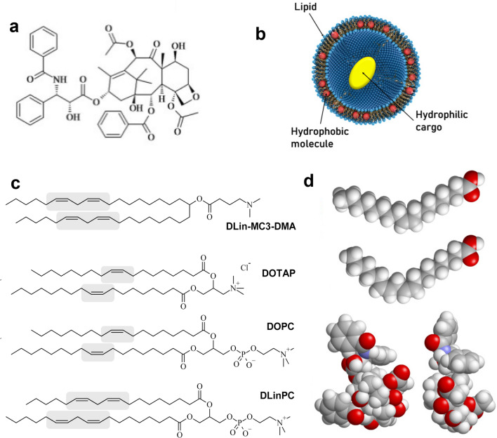
Figure 1.
Paclitaxel (PTX) and lipid vectors. (a) Chemical structure of PTX. (b) A unilamellar liposome consisting of a self-assembly of amphiphilic lipid molecules. The liposome can carry hydrophobic molecules (red spheres) within its hydrophobic bilayer and hydrophilic molecules (yellow oval) in its aqueous interior. (c) Chemical Structures of DLin-MC3-DMA (the cationic lipid used in patisiran), DOTAP, DOPC (the lipids used in the Endo-TAG formulation of PTX) and DLinPC. The cis double bonds in the lipid tails are highlighted. (d) Space filling molecular models of the ground state structure of oleic acid (C18:1) and linoleic acid (C18:2) together with two views of the structure of PTX for size comparison. The structures were rendered using RasTop 2.2 (https://www.geneinfinity.org/rastop/).

