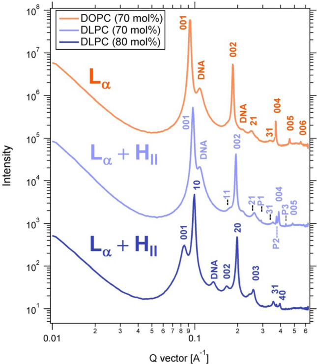
Figure 5.

X-ray scattering profiles of CL–DNA complexes prepared from PTX-loaded CLs with high contents of DOPC or DLinPC, revealing their self-assembled structures. Peak assignments shown are for LαC (00L), HIIC (HK), DNA–DNA interaxial spacing in the LαC phase (DNA), and crystallized PTX (P1, P2, P3). The CLs were composed of 70 or 80 mol% neutral lipid (DOPC or DLinPC), 2 mol% PTX, and the remainder DOTAP and were complexed with calf thymus DNA at a 1:1 charge ratio.
