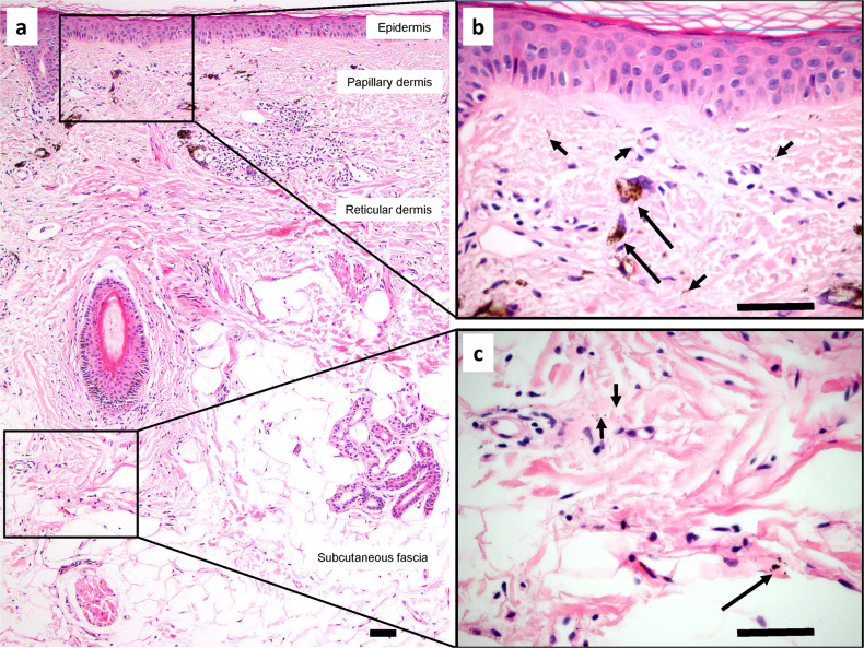
Fig. 1. Tattoo pigment in interstitial spaces of the dermis and subcutaneous fascia.
a H&E section of skin and subcutaneous fascia with cosmetically injected brown-black tattoo pigment. Pigment particles are present in the papillary and reticular dermis and subcutaneous fascia, visible at low magnification. b, c Higher magnification views of the rectangular areas demonstrate both intracellular particles (within macrophages; long arrows) and extracellular particles (within interstitial spaces; short arrows) of the papillary dermis (b), reticular dermis, and subcutaneous fascia (c). Scale bars = 100 μm.

