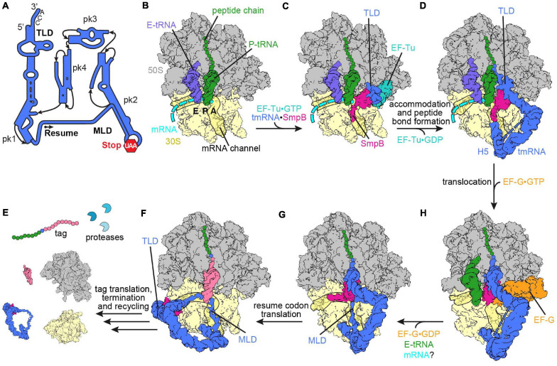
FIGURE 2.
Ribosome rescue by trans-translation. (A) Schematic representation of tmRNA (pk, pseudoknot; TLD, tRNA-like domain; MLD, mRNA-like domain). The arrow marks the resume codon and the red hexagon indicates the stop codon (UAA) of the MLD. (B) Non-stop complex (large subunit, 50S, gray; small subunit, 30S, yellow) with peptidyl-tRNA (green) in the P-site, E-site tRNA (slate blue) and mRNA (cyan) [PDB ID 4V8Q (Neubauer et al., 2012)]. (C) Delivery of the tmRNA-TLD (blue) in complex with SmpB (violet red) to the non-stop complex by EF-Tu (light sea green) [PDB ID 4V8Q (Neubauer et al., 2012)]. (D) tmRNA⋅SmpB (blue and violet red, respectively) accommodated to the A-site of a non-stop complex [PDB ID 6Q97 (Rae et al., 2019)]. Helix 5 (H5) of tmRNA binds close to the mRNA entry channel. The flexible MLD is indicated by the dashed line. (E) Post-translocation intermediate state of tmRNA⋅SmpB with EF-G (orange) [PDB ID 4V6T (Ramrath et al., 2012)]. tmRNA⋅SmpB and the tRNA (green) are in ap/P and pe/E hybrid states, respectively. (F) Post-translocation complex with tmRNA⋅SmpB in the P-site [PDB ID 6Q98 (Rae et al., 2019)]. The C-terminal tail of SmpB occupies the E-site of the mRNA channel and the resume codon is placed in the A-site. (G) Translation of the resume codon has occurred and the peptide chain was transferred from the TLD to the tRNA (pale violet red) decoding the resume codon [PDB ID 6Q9A (Rae et al., 2019)]. The TLD and SmpB are past the E-site at the outside of the ribosome, while the MLD is fully loaded to the mRNA channel. (H) Translation of the tag peptide, termination at the MLD stop codon and subsequent ribosome recycling have occurred. The tagged peptide is targeted by proteases.

