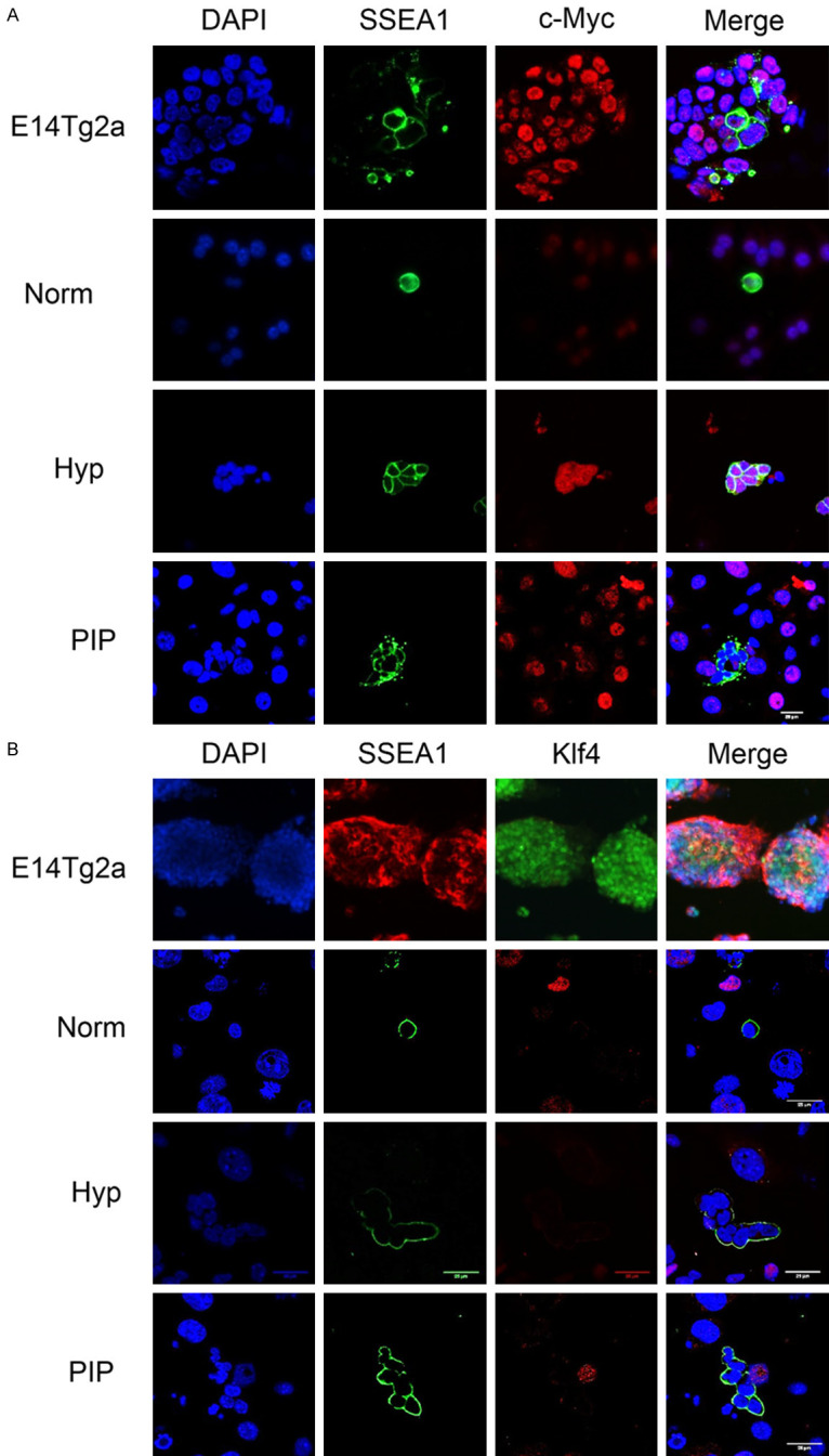
Figure 7.

Immunofluorescence microscopy against SSEA1 and c-Myc (A) and Klf4 (B) in PGC cultures, showing a negative reaction in SSEA1+ cell (PGCs) nuclei in PIP-supplemented cultures. Images also show PGCs cultured in normoxia (norm) and hypoxia (Hyp) for 48 h. Positive controls are the ESC line E14Tg2 and also hypoxic PGC cultures are positive for cMyc. Scale bars correspond to 25 µm.
