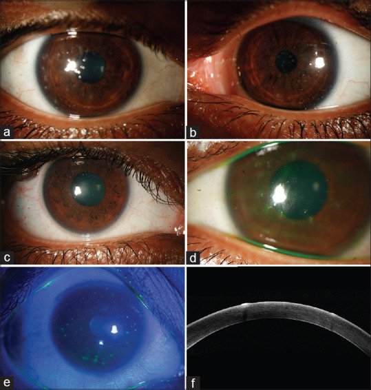
Figure 1.

Slit-lamp photograph showing: (a and b) multiple epithelial lesions in a patient with bilateral involvement, (c) multiple epithelial lesions, (d) fluorescein-stained epithelial lesions involving central cornea, (e) fluorescein-stained positive lesions seen under cobalt blue filter light, (f) anterior segment ocular coherence tomography showing hyperreflectivity of the epithelial lesion with back shadowing into the corresponding corneal stroma
