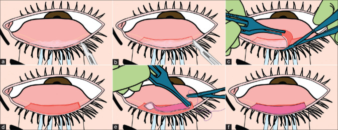
Figure 4.
Illustrations describing the surgical steps of lid margin mucous membrane grafting. (a) Everted and properly exposed keratinized lid margin of the upper lid. (b) Marking of a rectangular area including the keratinized lid margin and 4 mm of tarsal conjunctiva excluding 4-5 mm at medial and lateral ends. (c) Dissection of entire keratinized margin with tarsal conjunctiva off the tarsus by starting at one of the vertical edges. (d) The dissected bed is usually sized 18-20 mm horizontally and 4-5 mm vertically. (e) Suturing of labial mucosal graft from one end with 7 0 polyglactin sutures. (f) After completion of suturing, area of the bed should be larger than the area occupied by graft. A more detailed description of the surgical technique can be found at https://www.youtube.com/watch?v=SzCu-LbVlhs

