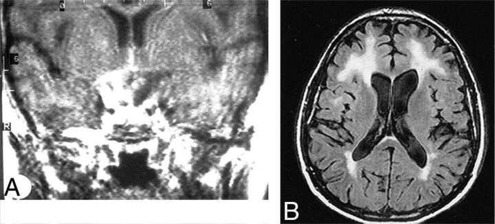
Fig 2.
Neurobrucellosis. Coronal postgadolinium MR imaging (A) shows sellar and suprasellar thickening and enhancement, presumed granuloma formation. Abnormalities resolved after treatment. Axial FLAIR (B) of a different patient shows extensive white matter hyperintensities in a 50-year-old patient. White matter changes in brucellosis may be due to demyelination, perhaps from autoimmune reaction. Meningeal enhancement and vascular changes are also reported in brucellosis. Reprinted with permission from Al-Sous et al.41

