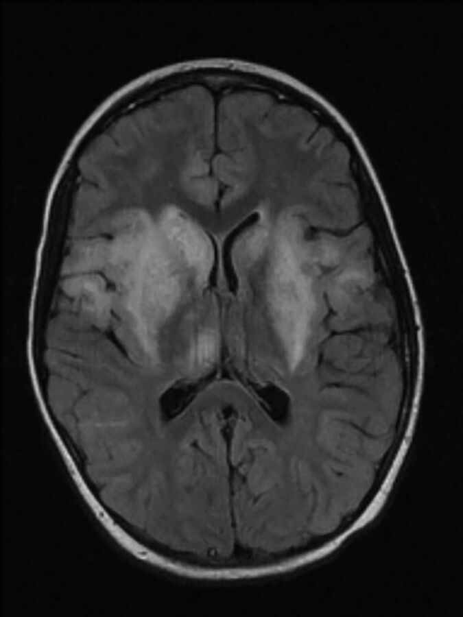
Fig 4.
Eastern equine encephalitis. FLAIR image illustrates basal ganglia involvement typical in this disease and involvement of the thalami, insula, and more peripheral cortex. Reprinted with kind permission from Springer Science and Business Media from Lury and Castillo.51

