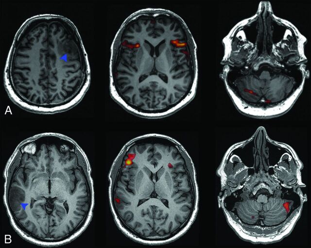
Fig 3.
A, Language activation of a left-handed, bilingual patient, with tumor in the left middle frontal gyrus (blue arrowhead), showing symmetric cerebral activation and right-lateralized cerebellar activation. B, Language activation of a right-handed patient with tumor in the right middle temporal and angular gyri (blue arrowhead), showing atypical, right-lateralized cerebral activation and crossed cerebellar lateralization.

