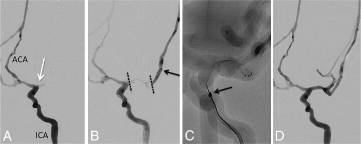
Fig 1.
Combined Stentriever plus aspiration approach to thrombectomy. A, Baseline angiogram, anteroposterior view, left ICA injection, demonstrating occlusion at the MCA origin (white arrow). ACA indicates anterior cerebral artery. B, Combined left ICA and microcatheter injection shows the extent of the thrombus within the left MCA (dotted black lines). Black arrow points to the tip of the microcatheter within an M2 branch of the MCA. C, Solitaire FR stent retriever is deployed within the M1 MCA segment. Black arrow points to the distal tip of the 5MAX ACE reperfusion catheter. D, Final angiogram after successful thrombectomy shows robust filling of the left MCA branches except for some distal superior trunk vessels. The final recanalization grade is TICI 2b.

