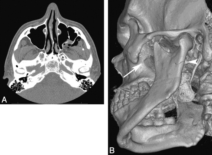
Fig 1.
A, A comminuted fracture of the posterior wall of the left maxillary sinus (arrow). Note the vector of the fracture fragment displacement (anteromedially) and herniation of the retroantral fat into the maxillary sinus. B, 3D reconstructed image demonstrates a minimally displaced fracture of the left subcondylar mandibular ramus, extending into the mandibular notch (arrowhead). The comminuted left posterior maxillary wall fracture is redemonstrated (long arrow).

