Fig 2.
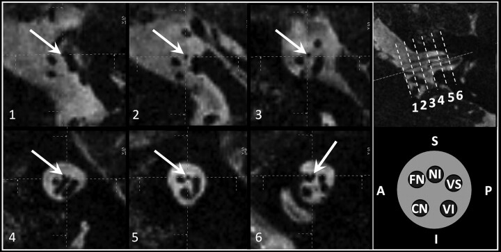
Parasagittal reformatted CISS sequence images (1–6) (TR, 12.18 ms; TE, 6.09 ms; flip angle, 50°) of the CPA and IAC at 3T depicting the course of the NI and additional reference images on the right: 1) NI originates (white arrows) anterior to the nervus vestibularis superior from the brain stem, 2) NI in the middle third of the CPA between the nervus facialis (anterior) and vestibularis superior (posterior), 3) NI in the distal third of the CPA between the nervus facialis (anterior) and vestibularis superior (posterior), (4) loop of the anterior inferior cerebellar artery in the proximal IAC slightly elevating the NI, (5) NI in the middle third of the IAC between the nervus facialis (anterior) and vestibularis superior (posterior), and 6) NI in the distal third of the IAC attached to the nervus facialis (anterior). Reference pictures: upper row, para-axial CISS sequence image with dotted reference lines for images; 1–6, lower row, schematic drawing of the order of nerves (clockwise direction) in the IAC.

