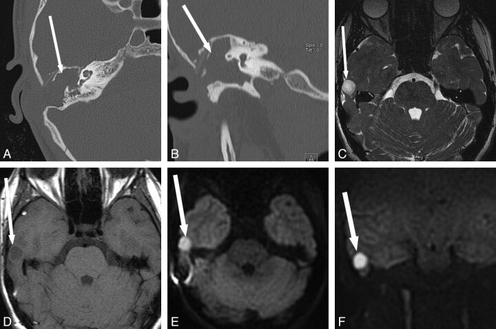
Fig 3.
Detection of recurrent disease and intracranial extension when otologic evaluation is obscured and CT is nonspecific. A 14-year-old girl with a long history of recurrent cholesteatoma and multiple surgeries. The middle ear is obscured due to a stenotic external auditory canal (A and B). CT shows nonspecific diffuse opacification of the mastoidectomy and middle ear (arrow). C−F, MR imaging shows T2 hyperintense (arrow, C) and T1 hypointense (arrow, D) regions with hyperintensity on BLADE DWI (arrow, E and F) along the superior aspect of the right temporal bone, suspicious for recurrent cholesteatoma with intracranial extension. The patient declined contrast material. At surgery, intradural extension of disease was confirmed.

