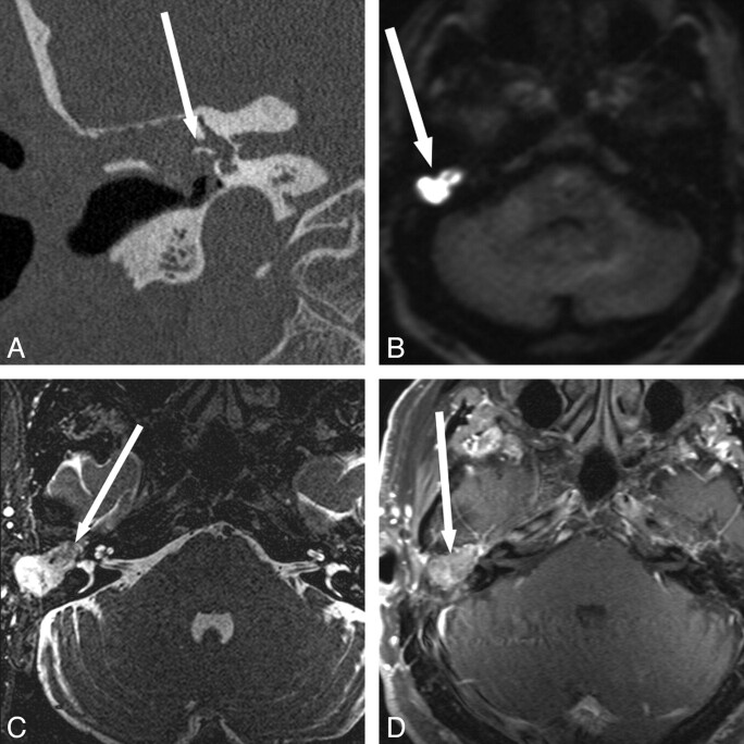
Fig 4.
Evaluation of disease extent in a patient with lateral semicircular canal fistula. A 61-year-old man with multiple prior ear surgeries, including a right mastoidectomy for unknown reasons, presented with vertigo and imbalance. Visual inspection of the middle ear cavities was obscured by postoperative changes, but no cholesteatoma was seen. A and B, CT shows soft tissue (arrow) in the mastoid defect, external auditory canal, and epitympanum with bony erosion of the lateral semicircular canal. C and D, MR images show the extent of cholesteatoma and demonstrate a large area of hyperintensity on HASTE DWI in the mastoid defect and middle ear (B in Fig 1) with T2 hypointensity (arrow, C), and mild T1 hyperintensity but no definite enhancement (arrow, D). A portion of the right lateral semicircular canal is obscured by the soft-tissue mass (C), again consistent with the fistula shown on CT. Cholesteatoma and lateral semicircular canal fistula were confirmed at surgery. Fig 4A, B, and C reproduced with permission from Ear, Nose & Throat Journal (Schwartz KM, Lane JI, Neff BA, et al. Diffusion-weighted imaging for cholesteatoma evaluation. 2010;89:E14-19).32

