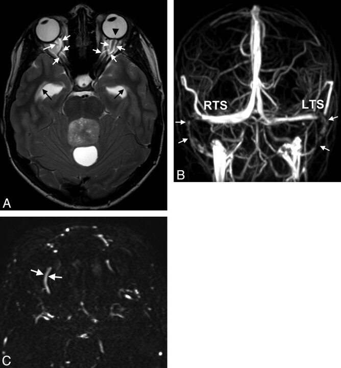
Fig 1.
A 15-year-old boy with double vision and reduced acuity has bilateral papilledema. A, On axial T2WI, ventriculomegaly with periventricular CSF flow (black arrows) due to an obstructing tumor within the fourth ventricle is found. Optic nerve sheath hydrops, elongation of the optic nerve (white arrows in A), and optic papilla protrusion are seen (black arrowhead). B, Anteroposterior view of MIP of MRV depicts bilateral narrowings of the lateral RTS and LTS and the sigmoid sinus (arrows). C, On primary axial sections of MRV, the SOVs are enlarged (arrows, right SOV, 2.8-mm width). Medulloblastoma was found at surgery.

