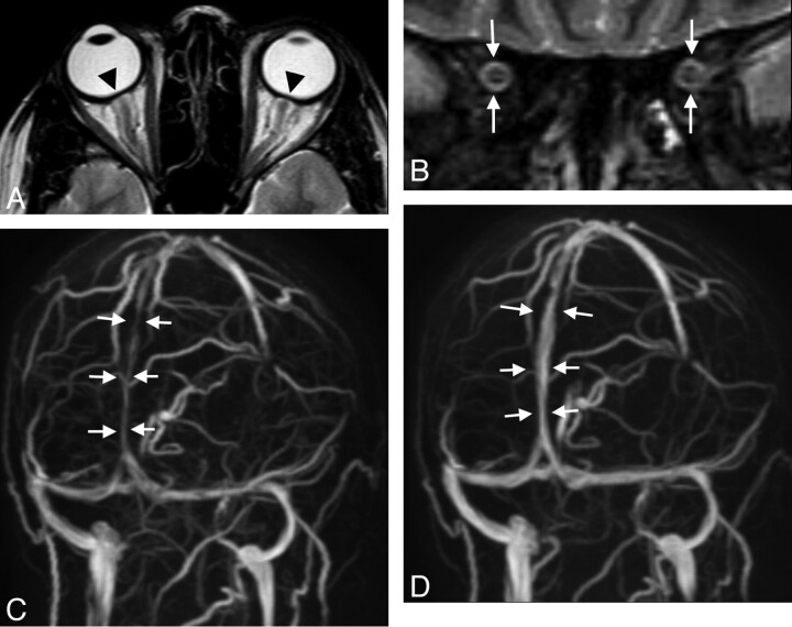
Fig 2.
A 30-year-old woman with arterial hypertension and visual disturbances has bilateral papilledema. A, MR imaging of the orbit displays a flattened posterior sclera (arrowheads). B, ONS hydrops is present, and there is edema of the optic nerve as seen on the coronal STIR sequence measured 20 mm behind the globe (white arrows, ONS width of 5.4 mm). C, Slightly oblique MIP of MRV shows lengthy narrowings of the intracranial venous sinuses, especially of the superior sagittal sinus (arrows). Vision returns to normal with successful treatment of hypertension. On follow-up MR imaging 3 months later, ONS width is reduced to 4.8 mm, and the posterior sclera appears normal (not shown). D, The intracranial venous sinuses regain normal caliber, as shown by MRV.

