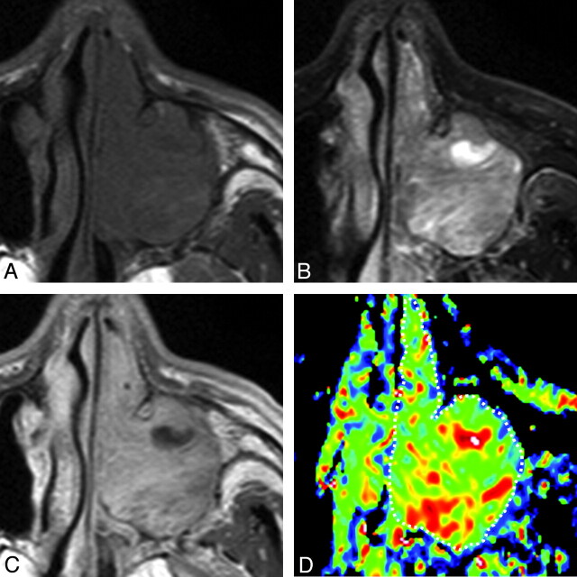
Fig 3.
A 69-year-old man with inverted papilloma. A, Axial T1-weighted MR image (TR/TE = 500/15 ms) shows tumor with homogeneous signal intensity filling the left maxillary sinus and nasal cavity. B, Axial FS (SPIR) T2-weighted MR image (TR/TE = 4784 ms/80 ms) shows the tumor with heterogeneous signal intensity. C, Axial CE T1-weighted MR image (TR/TE = 500/15 ms) shows heterogeneously enhanced tumor. D, Axial color ADC map shows tumor with scattered areas of high ADCs. Overall ADC = 1.4 × 10⁻³ mm2/s. Areas with extremely low, low, intermediate, and high ADCs occupy 0%, 21%, 66%, and 13%, respectively, of the tumor.

