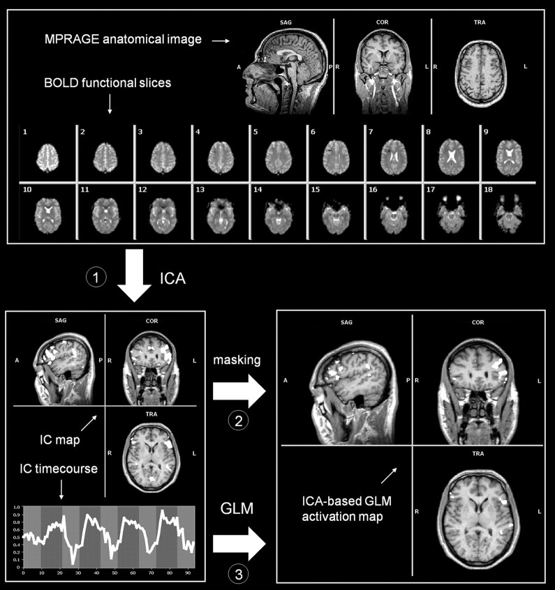
Fig 1.
Outline of the ICA-based GLM analysis. The procedure can be divided into 3 steps: 1) The fMRI time courses are decomposed by means of spatial ICA, and the IC showing the largest correspondence with the predictor is selected; 2) a spatial mask is created by thresholding the IC map, which is applied to the fMRI data; and 3) GLM analysis is performed on the masked fMRI time course as brain response data, by using the IC model.

