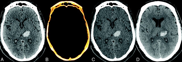
Fig 2.
Left thalamic intraparenchymal hyperattenuation due to hemorrhage of uncertain etiology in a 51-year-old man referred for altered mental status. The patient underwent a dual energy CTA and MR imaging for further evaluation. A, SE image shows left thalamic intraparenchymal hyperattenuation without corresponding hyperattenuation on the iodine overlay image (B). C, The focus of hyperattenuation is well demonstrated on the VNC image. A 24-hour follow-up NCCT scan (D) demonstrates largely stable hyperattenuation in the left thalamus, with an increase in the surrounding edema, confirming the original diagnosis of intraparenchymal hemorrhage.

