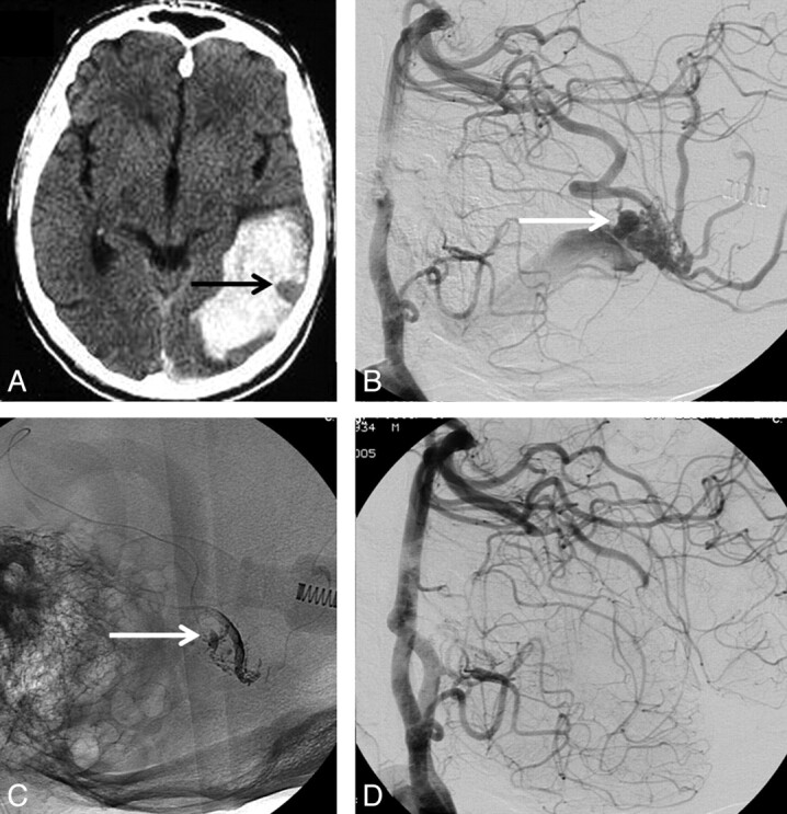
Fig 1.

71-year-old man with sudden headache and hemianopsia. A, CT scan shows posterior temporal hematoma with an outlined venous aneurysm indicating the source of the hemorrhage (arrow). B, Lateral view of vertebral angiography shows micro-AVM with intranidal aneurysm as the source of hemorrhage (arrow). C, Nonsubtracted lateral view shows Onyx cast after embolization. The intranidal aneurysm is partly filled with Onyx (arrow). D, Complete obliteration of the AVM with Onyx.
