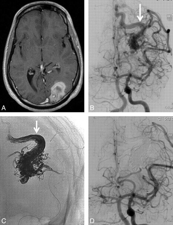
Fig 3.
23-year-old man with sudden headache and hemianopsia. A, T1-weighted MR imaging demonstrates left occipital hematoma. B, Frontal view of concomitant left internal carotid and vertebral angiogram shows small occipital AVM with a single draining vein (arrow). C, Nonsubtracted image of Onyx cast after embolization: complete filling of the nidus and proximal part of the draining vein (arrow). D, Angiography demonstrating complete obliteration of the AVM.

