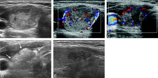
Fig 2.
An example of successful US-guided percutaneous EA for a circumferential peripherally located solid component remaining in a 34-year-old woman following RFA. A and B, Transverse gray-scale and color Doppler US images of a thyroid nodule (1.5 × 2.3 × 2.4 cm) in the right lobe show isoechogenicity and mildly increased vascularity, respectively. C, Transverse color Doppler follow-up US image obtained 6 months after the RFA session shows mild shrinkage (1.1 × 1.5 × 1.5 cm, decrease in volume of 70.1%) and the presence of vascularized solid components in the peripheral regions of the nodule. D, Transverse gray-scale US images just after EA show complete replacement of the nodule with intranodular echo staining (arrows) due to injected ethanol (total amount of injected ethanol, 1 mL). E, Longitudinal gray-scale follow-up US image obtained 6 months after a single session of EA shows marked hypoechogenicity and no vascularity (0.5 × 0.8 × 1.1 cm, decrease in volume of 94.7%).

