
Figure 1.
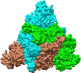
Inter‐subunit contacts between RBDs are maximized in the locked spike. This top view of the SARS‐CoV‐2 spike trimer with three linoleate molecules bound following 200 ns of MD simulation of the 6ZB5 PDB structure, illustrates the close contacts between the subunits. Individual chains are shown in surface representation and are colored cyan, green and brown.