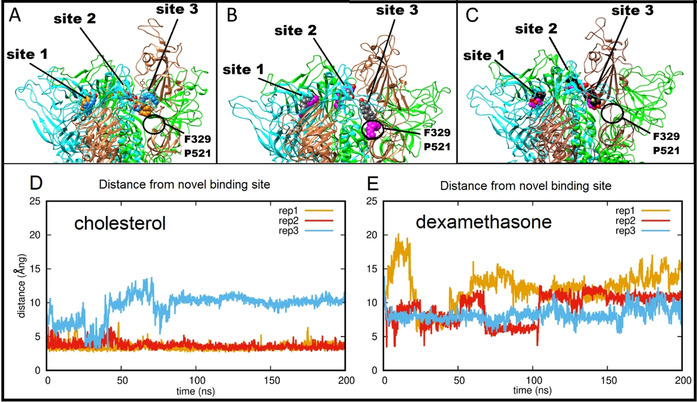
Figure 5.

Ligand interactions of the SARS‐CoV‐2 spike protein in the open form. A) Starting (carbon atoms shown in orange) and final (carbon atoms blue) positions of three LA− molecules from one 200 ns MD simulation of their complex with the SARS‐CoV‐2 spike in the open conformation. B) Starting (carbon atoms grey) and final (carbon atoms magenta) positions of three cholesterol molecules from an equivalent 200 ns MD trajectory. C) Starting (carbon atoms black) and final (carbon atoms purple) positions of three dexamethasone molecules in the last frame of an equivalent 200 ns MD trajectory. A black circle (panels A, B, C) denotes the alternative binding site 3 sampled by the site 3 cholesterol (see main text). D, E) Time dependence of distance between the site 3 cholesterol (D) or dexamethasone (E) and residues F329 and P521 of the alternative site 3 favored by cholesterol in the hinge region.
