
Figure 6.
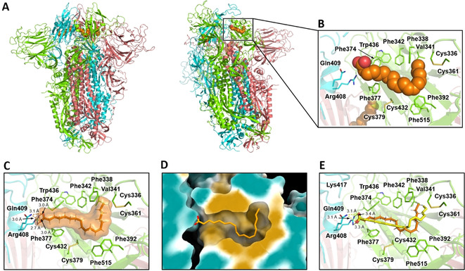
BUDE docks linoleate into the locked SARS‐CoV‐2 spike trimer in a conformation consistent with cryo‐EM structures. A) Cryo‐EM structure of the LA−‐bound spike trimer in the locked conformation. B) LA− binding site in the same structure. C) LA− binding pose in the structure used for docking (i.e. the cryo‐EM structure after MD equilibration). D) Surface representation of the LA− binding pocket in this structure, colored according to hydrophobicity: cyan for most hydrophilic through white to gold for most hydrophobic. The linoleate binding mode after equilibration by MD is shown as sticks. E) Superimposition of the BUDE predicted linoleate binding pose (yellow) and MD equilibrated structure (orange); the RMSD between the two poses is 1.6 Å.