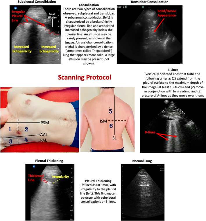
Figure 1.

Scanning protocol and lung ultrasound findings in COVID‐19 patients. This study utilized a 12‐zone protocol. 27 On each hemithorax, there are 6 zones. The exam begins on the patient's right side. Zones 1–2 (anterior zones) are between the parasternal margin (PSM) and the anterior axillary line (AAL) and are best obtained in the mid‐clavicular line. Zones 3–4 (lateral zones) are between the AAL and posterior axillary line (PAL) and are best obtained in the mid‐axillary line. The nipple line serves as a bisecting area between these zones. Zones 5–6 (posterior zones) are medial to the scapular line (SL) and are bisected by the inferior scapular margin (ISM). The zone areas are repeated on the contralateral hemithorax (starting with zone 7). This figure contains an overview of the observed ultrasound findings based on previously described terminology. 3 , 27 , 28
