TABLE 2.
Chest X‐ray

|

|

|
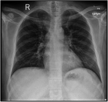
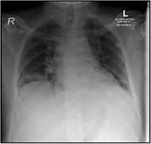
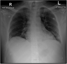
| At Admission: An increase in bronchovascular markings in the mid and lower zones. The transverse lines in the lower zone of the right lung may indicate the plate atelectasis. There is also suspicion of very fine shadowing in the periphery of both lower zones, and therefore, any parenchymal infiltrates cannot be ruled out. Close follow‐up is recommended | At development of pain crisis and acute chest syndrome 21/11/2020: Interval progression of bilateral basal atelectasis and faint infiltrates more on the right side | 23/11/2020: XR Chest incomplete resolution of the widespread consolidation distributed over both lung fields when compared to a chest X‐ray done 2 days ago the condition is improving |
