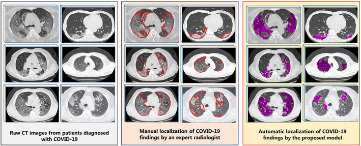
FIGURE 8.

Automatic localization of COVID‐19 pneumonia findings with the model proposed: Raw computed tomography (CT) images without any processing, manual screening of COVID‐19 findings by an expert radiologist (red borders), visualization of the findings by automatically scanning the same images with the proposed model (the findings are represented in purple) [Color figure can be viewed at wileyonlinelibrary.com]
