
FIGURE 1.
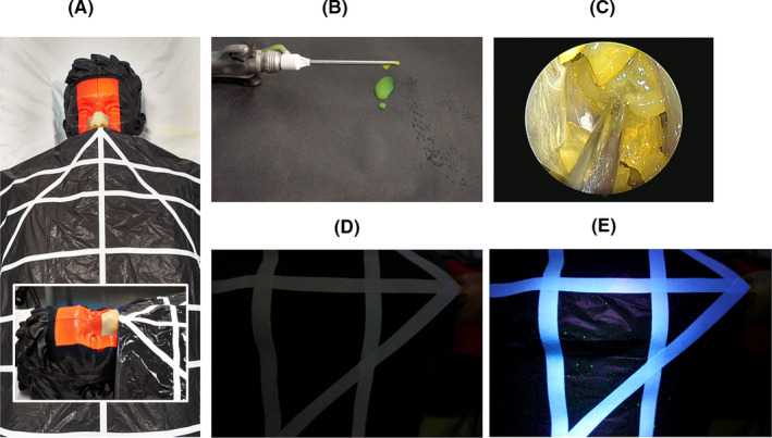
Experimental setup: A, Model of the head draped with grid detection sheet. Inset shows close‐up of the 3‐D printed nose and paranasal cavity. B, Example of dripping from microdebrider after activation. C, Endoscopic view of fluorescein stained grapes mimicking nasal polyps. D,E, Example of droplets identified on detection grip before and after UV lamp illumination, respectively