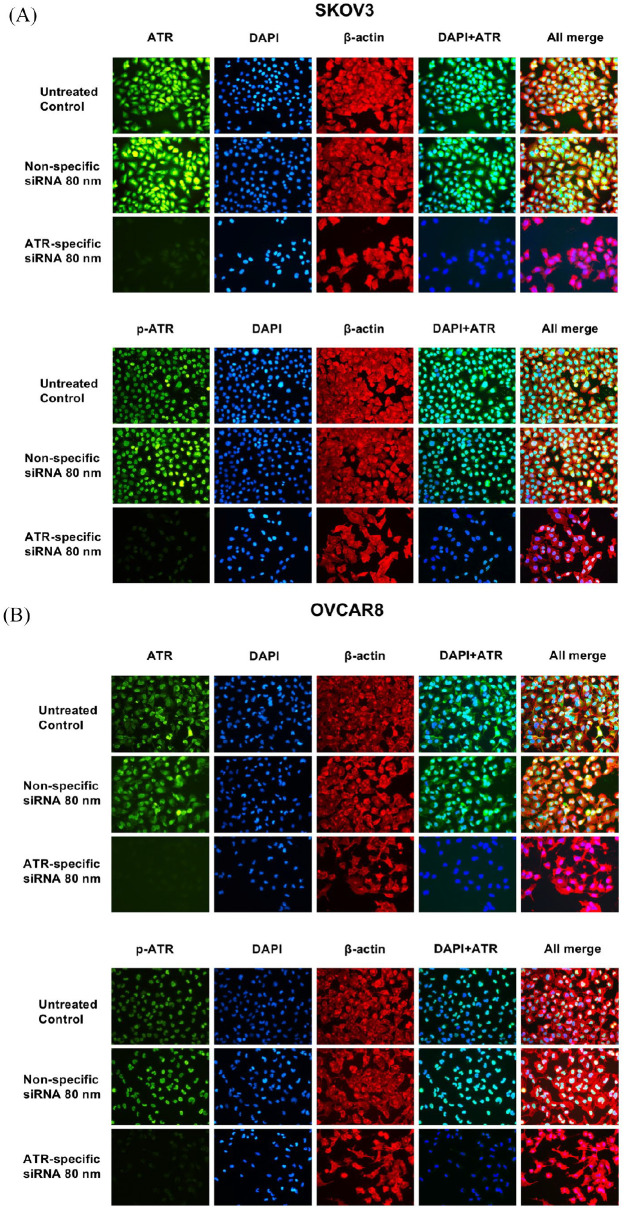
Figure 5.
ATR and p-ATR expression in SKOV3 (A) and OVCAR8 (B) ovarian cancer cell lines was assessed by immunofluorescence with antibodies to ATR (green), p-ATR (green) and β-actin (red). Hoechst 33342 was added to counterstain the cell nucleus (blue). Green fluorescence of ATR resided within the cytoplasm and nucleus, whereas p-ATR protein was localized mainly in the nucleus alone. Expression of ATR, p-ATR, and cell proliferation were significantly reduced by ATR-specific siRNA treatment compared with non-specific siRNA treatment.
ATR, ataxia-telangiectasia and Rad3 related; DAPI, 4′,6-diamidino-2-phenylindole; p-ATR, phospho-ATR ser428.

