
Figure 1.
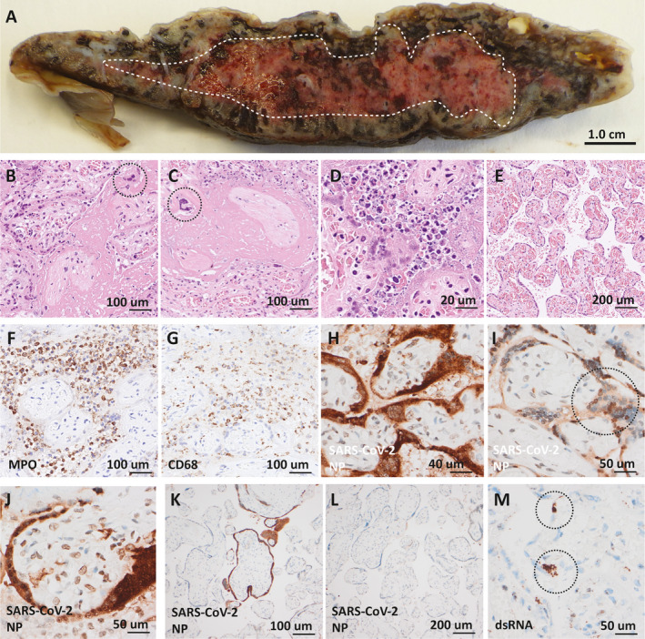
Placental pathology. (A) Transected placenta with confluent accumulation of fibrinoid demarcated (white broken line). (B,C) Massive intervillous fibrinoid deposition surrounding denuded villi with extravillous syncytiotrophoblasts (circles) located in the fibrinoid. (D) Acute intervillositis with karyorrhectic neutrophils in the intervillous space and degeneration of the villous trophoblast layer. (E) Representative region of chorangiosis. (F,G) Immunohistochemical staining for myeloperoxidase (MPO) and CD68 with positivity in inflammatory cells in areas of intervillositis. (H‐J) Severe acute respiratory syndrome coronavirus‐2 (SARSCoV‐2) nucleoprotein (NP) detected in nucleus and (circle in I) and cytoplasm in villous trophoblasts and syncytiotrophoblasts as well as in the nucleus of villous stromal cells (J) in areas of intervillositis. (K,L) Areas without intervillositis showed absent or focal staining for SARS‐CoV‐2 nucleoprotein of villi. (M) Double‐stranded RNA (dsRNA) detected in villous trophoblasts and syncytiotrophoblasts (circles).