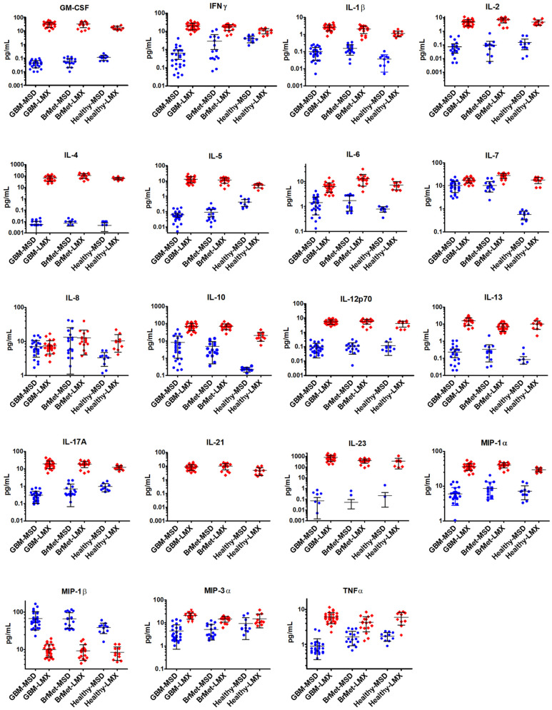
Figure 3.
Cytokine concentrations in human plasma samples. All 19 shared cytokine analytes were evaluated in 55 human plasma samples comprising 27 glioma subjects, 17 subjects with secondary brain metastases (BrMet), and 11 healthy controls (HC). Values shown are mean concentration ± SD. The Luminex (LMX) platform categorized identical samples at greater concentrations than the Meso Scale Discovery (MSD) system for most analytes. With IL-4, wherein LMX categorized cytokine concentrations at 4 orders of magnitude higher than MSD, and GM-CSF values at 2-3 orders of magnitude greater than MSD values. Notably, with IL-21, although the MSD lower quantification limit was 0.22 pg/mL, none was detectable by MSD in any sample. The LMX platform identified IL-21 in all samples (range 1.8-18.9 pg/mL). With IL-23, the LLoQ was 55-fold more sensitive using MSD (0.22 pg/mL) than LMX (12.15 pg/mL). While IL-23 was quantifiable by LMX in all samples, MSD detected the analyte in only 8% of samples, and categorized them at lower concentrations (range 0.24-2.03 pg/mL) than LMX (range 86.8-1922.2 pg/mL). The MSD platform quantified 7 cytokines in 100% of samples (IL-6, IL-7, IL-8, IL-10, MIP-1β, MIP-3α, and TNF-α), and expression trends of each of these across the 3 experimental groups were generally similar between MSD and LMX. With MSD, IL-7 levels appeared lower in HC samples versus GBM and BrMet, which was not observed with LMX assessment. While IL-17A was detected in 100% of samples by LMX and 76% by MSD, there a contradictory trend appeared in which IL-17 was lower in HC versus the 2 neoplasm groups with LMX, but higher with MSD. Only MIP-1β levels were categorized as greater using MSD (range 16.06-166.88 pg/mL) versus LMX (range 4.26-19.65 pg/mL).

