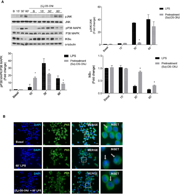
Figure 3.
Effects of (S S)-DS-ONJ in the activation of MAPKs and NFκB-mediated signaling in LPS-stimulated Bv.2 microglial cells. Bv.2 microglial cells were stimulated with 200 ng/mL LPS in the absence, or presence, of 10 μM (S S)-DS-ONJ for the indicated time periods. (A) Protein extracts were separated by SDS-PAGE, and analyzed by Western blot with antibodies against phosphorylated (p)-JNK, total JNK, phosphorylated (p)-p38α MAPK, total p38α MAPK, IκBα and α-tubulin. Representative autoradiograms are shown (n = 7 independent experiments). Blots were quantified with scanning densitometry, and the results are presented as mean ± SEM. The ratios between the indicated proteins and the fold changes relative to the basal values are shown. *p ≤0.05 vs LPS treatment (two-way ANOVA followed by Bonferroni t-test). (B) Confocal immunofluorescence assessment of the nuclear translocation of p65-NFκB in Bv.2 microglial cells following stimulation with LPS in the presence or absence of (S S)-DS-ONJ. Activation of p65-NFκB nuclear translocation was defined by an increase in immunofluorescence of p65-NFκB (green channel) in the nuclear regions. Nuclear regions of Bv.2 microglial cells were determined by counterstaining nuclear DNA with DAPI (blue channel). White arrows indicate the p65-NFκB nuclear o cytoplasmic localization.

