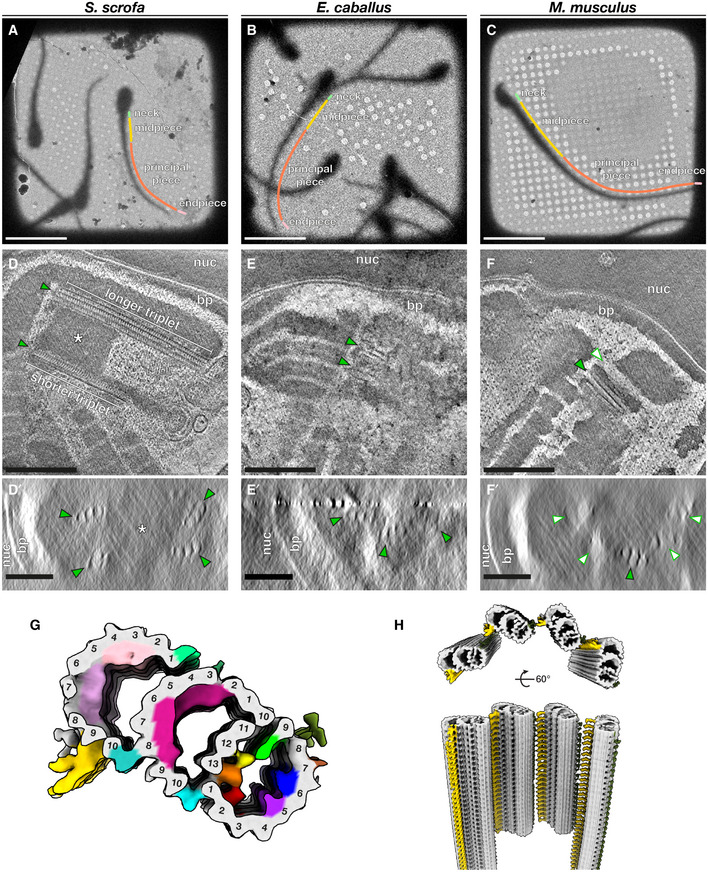
Figure 1. The proximal centriole (PC) in mammalian sperm is asymmetric and contains novel microtubule inner proteins.

-
A–CLow‐magnification cryo‐EM projection images of pig (A), horse (B), and mouse (C) sperm. Different regions of the flagellum discussed in this paper are annotated as follows: green—neck, yellow—midpiece, coral—principal piece, pink—endpiece.
-
D–FTomographic slices through cryo‐FIB‐milled lamellae of pig (D), horse (E), and mouse (F) sperm. Transverse slices (D’–F’) show complete triplets in the pig (D’) and the horse (E’), but not in the mouse (F’). Complete triplets are indicated by green arrowheads with black outlines, while degenerated triplets are indicated by white arrowheads with green outlines. Note the electron‐dense material in the lumen of the pig sperm proximal centriole that is continuous with the connecting piece (asterisks in D and D’).
-
GIn situ structure of triplet microtubules from the proximal region of the pig sperm PC with the tubulin backbone in gray and microtubule inner protein densities colored individually. A‐tubule MIPs are colored: MIP1—green, MIP2—yellow, MIP3—orange, MIP4—red, MIP5—purple, MIP6—blue. B‐tubule MIPs are colored: MIP7—magenta. C‐tubule MIPs are colored: MIP8—light pink, MIP9—pink. The A‐C linker is colored gold and the putative A‐link is colored olive green.
-
HReconstruction of the proximal region of the pig sperm PC generated by plotting the average back into the original particle positions and orientations in the tomogram. This plotback only contains four triplets as only part of the centriole was captured in the lamella. The A–C linker is colored in gold and the putative A‐link in olive green.
Data information: Labels: nuc—nucleus, bp—baseplate. Scale bars: (A–C) 20 µm; (D–F) 250 nm; (D’–F’) 100 nm.
